WELL WE FOUND OUT THAT FOR SOME UNKNOWN REASON, MARKS SURGERY HAS BEEN CHANGED TO FEBRUARY 10th. SO WE GO FOR PRE-OP. ON THE 9th AND THE SURGERY THE DAY AFTER. KEEP US IN YOUR THOUGHTS AND PRAYERS.
THE DOCTOR ASSURED ME, MARK IS AS HEALTHY AS POSSIBLE AND THIS IS THE KIND OF CASE HE LIKES TO TAKE ON. MARKS CHANCES OF HAVING A SUCCESSFUL SURGERY ARE "98%"! OPPOSED TO 50%. I ALSO UNDERSTAND ANY TIME YOU GO INTO ANY SURGERY YOUR WALKING INTO A SNAKES DEN (AS DR. LEVI) PUT IT. BUT I AM CONFIDENT IN THE SURGEON DR.BRIAN REEMTSEN, FURTHER MORE THE STAFF AT UCLA IS SOME OF THE FINEST PEOPLE, AND ARE WORTHY OF ENTRUSTING THE CARE OR MY SON TO.
Tuesday, January 27, 2009
Monday, January 26, 2009
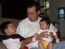
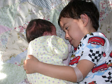
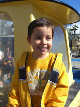
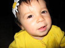
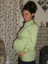
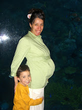
Pictures of Mark
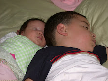
 Mark was only 2 or 3 days old
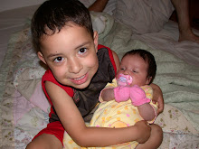
Marky 1st birthday

Mark at age 2

Marky at Legoland 2008 at Age 3

I love this pic. it was taken in Circus Circus, Las Vagas
THIS IS AN EXPLANTION ABOUT MARK'S HEART
I was Searching the web, for a good explantion to put on the blog, and I found one. I want you to understand a little about Mark-Anthony's Heart. I know this is a lenthie explantion but here it is...
Tricuspid Atresia (TA) is an abnormality of the heart valve and right side of the heart that occurs to a baby’s heart before birth. The normal opening between the right atrium (upper chamber of the heart) and the right ventricle (lower chamber of the heart) is called the tricuspid valve. When the tricuspid valve does not form during a baby’s development before it is born, blood is unable to flow between the two chambers. With the absence of this blood flow, the right lower chamber does not develop into a normal size. Blood is then forced to travel from the right upper chamber into the left upper chamber through a hole called the patent foramen ovale (PFO) a normal opening between the right and left upper chamber in the baby’s heart before it is born. After birth this hole should close on its own, but does not in this defect since the blood flow keeps it open. This opening allows blood that has been oxygenated by the lungs (red blood) to mix with the unoxygenated blood (blue blood) from the right upper chamber. This mixing causes the blood to have a lower oxygen content overall. This mixed blood is pumped into the left lower chamber. If there is a hole (ventricular septal defect) between the right and left lower chambers, then the mixed blood will travel to the lungs, through the small right lower chamber and the main vessel to the lungs (the Pulmonary Artery) as well as to the body via the left lower chamber and the main blood vessel to the body (the Aorta). Once the baby is born, it has the normal need for oxygen and relies on the lungs to perform the normal exchange of carbon dioxide and oxygen and to give the blood oxygen (red blood). Infants born with Tricuspid Atresia that do not have a hole between the two lower chambers have a small amount of blood flow to the lungs, provided by another small blood vessel called a patent ductus arteriosus (PDA). The PDA normally closes within a few hours up to a few days after the baby is born. This lack of red blood causes the baby to become slightly blue or cyanotic. The baby’s heart will beat faster to pump more blood to the lungs and the baby will begin to breathe faster trying to take in more air. The more work the heart and lungs do to increase the amount of red blood to the body, the more need there is for more red blood. This cycle will cause the baby’s body to release chemicals into the blood that will eventually cause the baby’s heart to stop beating. This could take as long as two weeks to happen if the heart defect is not found.
To fix the heart with this defect usually takes three operations, with the first one as soon as possible after the defect is discovered. If there is a hole between the two lower chambers (ventricular septal defect), but without any narrowing of the pulmonary artery, a small band is placed around the pulmonary artery as a temporary procedure. This temporary procedure prevents too much blood from flowing to the lungs. If there is not enough blood flow to the lungs then a shunt is created, the shunt connects the blood flow from the aorta to the pulmonary artery to supply the lungs with blood. The size of the shunt is based on the infant’s size. The second operation is the Glenn anastomosis and is done when the infant is between four and six months of age. The superior vena cava is sewn to the right pulmonary artery increasing the blue blood flow to the lungs. The shunt is removed at this time. The final operation is the Fontan Procedure. The inferior vena cava is sewn to the junction of the right pulmonary artery and superior vena cava. A special fabric tube (Gore-Tex) is used to create a separation between the inferior vena cava and the heart.
The tissue (atrial septum) between the two upper chambers (right and left atriums) is removed. The heart now has a single atrium and a single ventricle and the blood flow to the lungs bypasses the heart completely. Long-term outlook for the children will be the basis for this study. Most children have reasonably good exercise tolerance during their daily lives
Thursday, January 22, 2009

I've decided to blog for this year. As some of you may know Mark-Anthony's Heart surgery is Feb. 10. I am also expecting a baby girl on April 17th. So there is alot going on. And as much as I would like to call every member of my Family and extended family and Friends, it's just not possible. So i will give updates, on Marky condition well in the hospital and after. Then soon after post pictures of of a sweet baby girl.
Love, Joy
Love, Joy
Subscribe to:
Posts (Atom)